
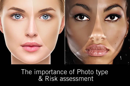
There have recently been several web forum and group discussions about photo type and relevance to skin analysis, and during these discussions there appeared to be for some participants, a rudimentary understanding of the importance of this characteristic in professional skin analysis. The purpose of this primer is to provide a good base of understanding. FBH
A key area of discussion should be about Skin Colour Terminology; and that we should clarify the reference to a skin colour and the burn time category within which it lays, by using the modern terminology of ‘photo type’ and risk. With the mixed ethnicities of today, the older Fitzpatrick Skin Types (Scale 1 to 6) terminology of skin colour analysis; although a good starting point, is no longer adequately applicative.
There was a controversy at the time that the Fitzpatrick Skin Type Scale was published in 1975 because still at that time there were light and dark skin tones within each race and the scale was considered too narrow to be useful. [1] However, that been said, the Fitzpatrick Skin Type Scale has been used ever since by the medical and cosmetic industries.
Nowadays there are many misunderstandings with the term ‘skin type’ used today; those of you trained in the Pastiche Method of skin analysis were taught that ‘skin type’ is a reference to the intrinsic skin groups of lipid dry, oily and diffused redness.
For others, the term ‘skin type’ often still refers to the original Fitzpatrick classification of skin colour; and today older medical professionals still make the reference of Fitzpatrick skin type or just skin type.
During the 1990’s the use of the Fitzpatrick Skin Type Scale was revised; however although the Scale was a good reference for the skins burn time, it did not give any indication of the skins ability to accumulate melanin and change colour (tan). Nor did it indicate the risk for skin cancer or post inflammatory hyperpigmentation.
Differences in each tone of skin colour can be seen more obviously in the Fitzpatrick Skin Type Scale of 4 to 6, and those differences associated with the genetics of hemisphere. (Location)
The closer to the equator, the darker the skin tone and when moving further north or away from the equator, the skin tone becomes lighter.
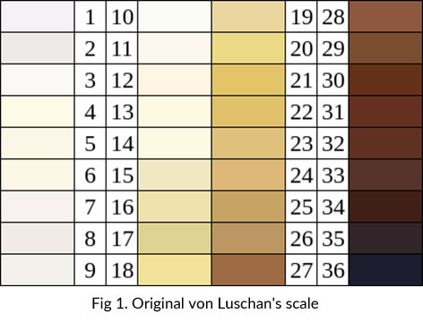
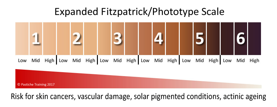
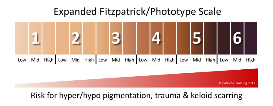
I use the very large continent India as a perfect example of this phenomenon. Therefore, because there are light and dark skin tones within each of the original Fitzpatrick Skin Type Scale, I have always recommended that each of the 6 types used in the scale is further broken down into a low, medium and high in order to add a higher resolution, or accuracy. This Photo Type Scale would be similar to the original von Luschan’s scale of 36 tones, but not so extensive. [2,3]
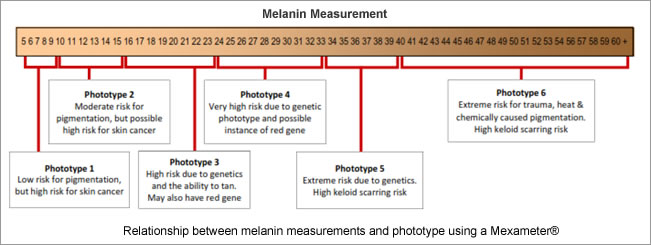
One of the reasons I did not allocate skin’s that I was analysing in to one of the 6 Fitzpatrick categories was that my skin diagnostic devices that measure melanin density in the skin were telling me that there were more variables in tone to allocate to a single type. In fact, some Skins that would normally be classified as one Fitzpatrick type could have fitted in to 9-sub tones according to the measurement scale. The diagram below illustrates the relationship. For example how would you accurately classify skin’s with readings of 23-25 or 32 to 35 on a conventional Fitzpatrick scale?
Understanding Skin response to UVR is important and separated into two categories;
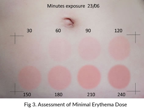
The first category is the term Minimal Erythema Dose; (acronym one MED). One MED was the point of reference used to describe the minimal exposure time to UVR to create visible redness (erythema) or for a skin colour to burn. It is commonly used today as an indication of a sunscreen’s Sun Protection Factor also known as the SPF.
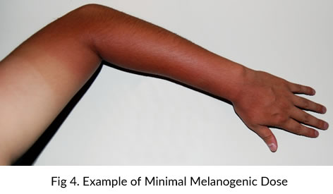
The other category is the Minimal Melanogenic Dose, (acronym MMD). We refer to MMD as the skin’s ability to tan or the epidermis’s ability to accumulate melanin. MMD is usually determined several days after UV exposure (immediate pigment darkening) and when a change in skin colour occurs several days later between exposed and non-exposed skin. (Delayed pigment darkening). MMD is the terminology used when describing the melanocytes ability to protect skin, and it has been proven that even the fairest skin can accumulate melanin within the epidermis over a period of a summer.
Understanding how skin responds to UVR exposure has high priority in the skin treatment professions today as is the correct terminology that enables us all to communicate at one level. To clarify and avoid misunderstanding, I suggest that the term Photo Type Scale becomes the standard reference as a way to classify the typical response of different colours of skin to ultraviolet exposure.
The Photo Type Scale can also indicate skin risk; this risk may be a genetic predisposition for skin cancer especially for those skins that carry the red hair gene. (MC1R) (Melanocortin 1 receptor)
Red hair appears most commonly in people with two copies of a recessive allele (Ref-4) on chromosome 16 which produces an altered version of the MC1R protein. The MC1R recessive variant gene that gives people red hair and non-tanning skin is also associated with freckles though it is not uncommon to see a redhead without freckles. Eighty percent of redheads have an MC1R gene variant, and the prevalence of these alleles is highest in Scotland and Ireland. (5)
Alternatively, the Photo Type Scale can indicate risk for post inflammatory hyperpigmentation, which affects photo types 4 to 6. The genetic risk for keloid scarring may also be summarized if the photo type was found to be in the 5 – 6 group.
Information gathered during the consultation can provide you with the genetic data to determine a skin that is at risk for either skin disorder. With the mixed ethnicities of this millennium, it is not always apparent that skin has the red hair gene and patients, or clients may not know their genetic heritage; It is important to know or determine this if the Photo Type is a high 3 or low 4.
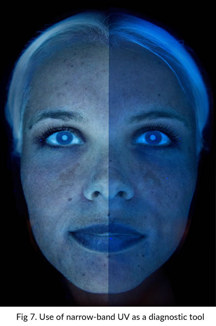
Diagnostic equipment is available now that help to determine photo type and measure accumulated melanin with great accuracy. [6] The modern, LED black light scanners show the hidden Ephelides (Freckles) that are an indicator of the red hair gene. [7] If the genetic history of a client shows a combination of the ability to tan and the red hair gene, (which is not uncommon among those of a photo type 3 and 4) it should be considered high risk for skin cancer and post inflammatory hyperpigmentation.
Because the ability to tan often means that the skin can tolerate a longer period of unprotected UV exposure, this combination of genes could culminate into skin cancer and pigmentation as the melanocyte and keratinocyte cells age and intrinsic antioxidant defense systems decline.
It was during my years as an Electrologist that I found that some of the lower Photo Types were prone to post inflammatory hyperpigmentation (PIH), not just the photo types 4 to 6 as indicated by the training of the day. It was after reviewing clients genetic history many years later that I attributed this susceptibility to PIH to the fact that many of my clients had mixed ethnicity’s and that their visible skin colour was not a reliable indication of their ‘risk’ for pigmentation.
This is another reason why the genetic heritage on both sides of the family tree is vitally important during a consultation.
The MMD was also a factor because if a skin measured as photo type 3 without sun exposure, I observed that the same skin could accumulate melanin and measure as a Photo Type 4 by the end of summer; thereby increasing the risk of post inflammatory hyperpigmentation during the wound healing process of electrolysis.
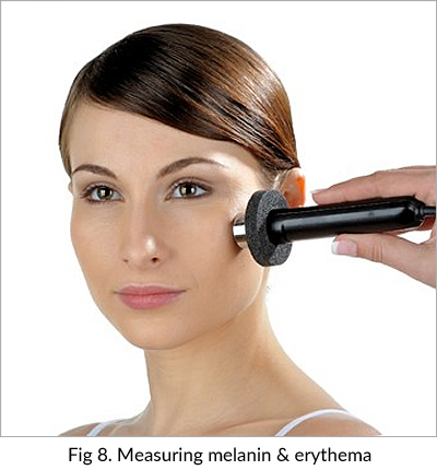
The modalities of intense pulsed light and laser commonly used for hair reduction and skin rejuvenation are taught with the emphasis on being mindful of accumulated melanin between treatments. Melanin measurements using equipment such as the CK SPA 99 [6] during the consultation and before each treatment ensure that an IPL/Laser devices fluence can be altered to accommodate any melanin increase or if appropriate, for treatment to be postponed.
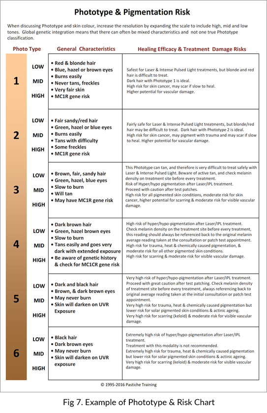
It was these experiences over time that prompted me to create a Photo Type Scale table that included the skin risk for modalities such as electrolysis, intense pulsed light, laser or collagen induction therapy. Today, this chart is used by educators and skin treatment professionals worldwide as a clinical reference tool. [8] See below.
References for this article
1.https://en.wikipedia.org/wiki/Color_terminology_for_race
2.“Felix von Luschan Skin Color chart” by C Burnett
3. https://en.wikipedia.org/wiki/Felix_von_Luschan
4.https://en.wikipedia.org/wiki/Allele
5.https://en.wikipedia.org/wiki/Red_hair
6.SPA 99 Skin pigmentation/Erythema measuring device
7. http://www.sylton.com/observ-
8.The A-Z of understanding pigmentation